File:Functional magnetic resonance imaging.jpg
Functional_magnetic_resonance_imaging.jpg (250 × 208 像素,檔案大細:11 KB ,MIME類型:image/jpeg)
檔案歷史
撳個日期/時間去睇響嗰個時間出現過嘅檔案。
| 日期/時間 | 縮圖 | 尺寸 | 用戶 | 註解 | |
|---|---|---|---|---|---|
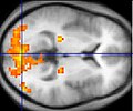
| 現時 | 2004年12月9號 (四) 00:52 |  | 250 × 208(11 KB) | Superborsuk | Sample fMRI data This example of fMRI data shows regions of activation including primary visual cortex (V1, BA17), extrastriate visual cortex and lateral geniculate body in a comparison between a task involving a complex moving visual stimulus and re |
檔案用途
以下嘅1版用到呢個檔:
全域檔案使用情況
下面嘅維基都用緊呢個檔案:
- ar.wikipedia.org嘅使用情況
- ast.wikipedia.org嘅使用情況
- az.wikipedia.org嘅使用情況
- bn.wikipedia.org嘅使用情況
- bs.wikipedia.org嘅使用情況
- ca.wikipedia.org嘅使用情況
- cs.wikipedia.org嘅使用情況
- da.wikipedia.org嘅使用情況
- de.wikipedia.org嘅使用情況
- el.wikipedia.org嘅使用情況
- en.wikipedia.org嘅使用情況
- Déjà vu
- Asperger syndrome
- Neurolinguistics
- User:Washington irving
- Functional neuroimaging
- Statistical parametric mapping
- Haemodynamic response
- Relapse
- Visual search
- Philosophy of mind
- Colour centre
- Neurolaw
- User:Letsgoridebikes
- Clinical neurochemistry
- User:Desoham3/Wikipedia Sandbox Color Center
- User:Hchandler52/sandbox
- User:Ironstamp/sandbox
- Neuroimaging intelligence testing
- Wikipedia:Top 25 Report/December 8 to 14, 2013
- User:Flyer22 Frozen/Human brain
- MRI pulse sequence
- User:ThunderhillMc/Déjà vu
- en.wikibooks.org嘅使用情況
- Cognitive Psychology and Cognitive Neuroscience/Behavioural and Neuroscience Methods
- Cognitive Psychology and Cognitive Neuroscience/Print version
- Chemical Sciences: A Manual for CSIR-UGC National Eligibility Test for Lectureship and JRF/Magnetic resonance imaging
- A-level Computing/AQA/Paper 2/Consequences of uses of computing/Emerging technologies
- A-level Computing 2009/AQA/Print version/Unit 2
- A-level Computing/AQA/Computer Components, The Stored Program Concept and the Internet/Consequences of Uses of Computing/Emerging technologies
- A-level Computing/AQA/Print version/Unit 2
- Lentis/Neuroprosthetics
- en.wikiversity.org嘅使用情況
睇呢個檔案嘅更多全域使用情況。
